
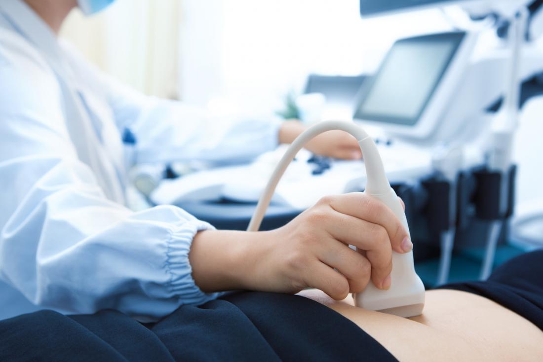
Ultrasound: how does it work?
Ultrasound takes its name from the Greek êchô , which means “sound” and graphy, or graphein in Greek, which means “to write”.
Ultrasound is one of the best-known imaging techniques. It is based on the properties of ultrasound, or sound waves, which are similar to vibrations, not audible to the human ear.
These ultrasounds are emitted in a beam by a probe and they spread out in a straight line. But their trajectory can be deviated when they reach a denser or more rigid medium. The encounter with these “obstacles” creates echoes which reflect the walls of the organs or the tissues. Using a computer, the echoes are converted into a cross-sectional and moving image of the region being examined. The morphology of the zone thus observable can then be analyzed.
This technique can also be associated with the use of a ” radar ” which allows the study of the vessels (doppler), as is the case for cardiac echo-doppler, for example.
When is an ultrasound performed?
Ultrasound is primarily used to visualize the morphology (size, shape, etc.) of organs, to know their evolution or good development, their location, etc.
This therefore makes it possible to carry out disease diagnoses , to ensure the monitoring of certain pathologies (evolution of cancers , endometriosis , etc.) or to guide the doctor in the context of certain surgical operations . We will not deal with this last aspect, which is more interventional, in this article.
Abdominopelvic ultrasound
The abdomino-pelvic ultrasound (or prostate if it concerns only the prostate) makes it possible to observe the organs of the abdomen and pelvis (uterus, ovaries, prostate and seminal vesicles or liver, gallbladder, kidneys and bladder).
It is useful for diagnosing pathologies that can affect these organs, such as the presence of ovarian cysts or endometriosis within the female reproductive system, or stones in the kidneys, or finally detecting infections of the gallbladder (cholecystitis) or kidneys for example (pyelonephritis).
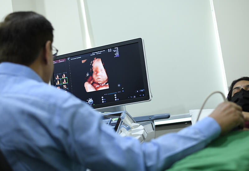
And it is also known to help monitor the pregnancy of pregnant women and ensure that the fetus is developing well and that there are no complications (miscarriage, ectopic pregnancy, etc.).
Two techniques are used. The organs can be observed through a probe placed on the skin of the belly. Ultrasound is called transcutaneous. In case of difficulties in benefiting from sufficiently clear and analyzable images, ultrasound is performed by endocavitary or endorectal route (for example for prostate ultrasound). The probe is then introduced inside the body (via the vagina or the rectum) to be as close as possible to the organs to be observed.
Cardiac echo-doppler
Cardiac echo-doppler is useful if there is a suspicion of cardiovascular disease, it makes it possible to observe several parts in motion. First of all, from the heart (cavities, valves…), but also from the vessels in charge of transporting blood to the heart and to the other organs of the body (this is the case of the aorta and the pulmonary arteries) and finally blood flow, to know how fast and in what direction the blood flows through the vessels. It is therefore used to diagnose diseases, malformations , heart failure ,arterial hypertension … It is also used to ensure follow -up after taking so-called “cardiotoxic” drugs.
It is performed by circulating a probe placed on the thorax and called “transthoracic” (ETT).
Kidney and bladder ultrasound
To explore the kidneys and the bladder, it is the kidney and bladder ultrasound that must be relied upon. It can be prescribed if the patient suffers from abdominal pain or if he is subject to urinary disorders (Presence of blood, pain). It can detect cysts, locate obstruction in the kidneys or stones for example.
Ultrasound in case of tumor
Finally, in the case of a tumor, ultrasound has the function of helping to differentiate between a cyst made up of liquid and a solid tumor, or even to know in which other areas or organs the cancer has developed. For example, thyroid cancer is asymptomatic before leading to potential lumps, difficulty breathing or swallowing. Ultrasound of the thyroid, making it possible to analyze its structure, the size of the gland, and the existence of nodules, is essential in case of doubt.
How is an ultrasound performed?
It can be practiced in many places (hospital, radiology practice, doctor’s office, etc.), as long as the room is sufficiently dark, for good visibility of the images. It does not last long, on average, 15 to 30 minutes and is ambulatory (no need to stay overnight). It can be carried out by a doctor specializing in the organ to be analyzed ( cardiologist , gynecologist, etc.), a radiologist or, in certain cases, by other health professionals.
Before the ultrasound, procedures may be required to make the organ easier to examine. In some cases, a laxative will need to be taken first. It will sometimes be necessary to come on an empty stomach, without drinking or eating, or on the contrary to drink large quantities of water.
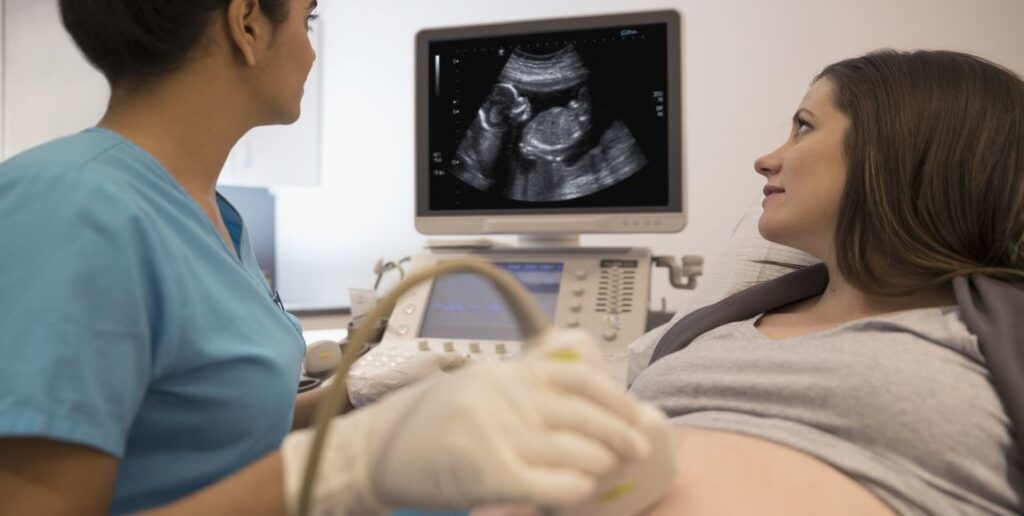
An assessment, relating to the medical history, current illnesses and medication taken, is carried out before the ultrasound. For the ultrasound itself, the patient is seated or lying down and may be asked to hold their breath. A probe, previously covered with an ultrasound-conducting gel, is positioned on the area to be analyzed (skin or by insertion into an orifice to be in direct contact with the organ). Connected to the computer, it makes it possible to transcribe moving images of the observed area using ultrasound. Some images are recorded so that the doctor can better analyze the morphology of the organs and in particular take measures. The results are generally communicated and explained after the examination, and formalized in a report, given to the patient and sometimes to the prescribing doctor.
If he deems it necessary, the doctor can recommend additional tests.

Ultrasound, is it painful?
Ultrasounds are not X-rays. The examination does not present any pain, danger or complications for the patient.
However, the pressure of the probe can be unpleasant if the area is weakened or in pain.
What documents to bring for an ultrasound?
If you are required to have an ultrasound, do not hesitate to ask your doctor’s office for the information you will need to provide on D-Day.
In particular, remember to bring your prescription, your carte Vitale, your mutual insurance card, and any other important element of your medical file (Letters from the doctor, results of examinations, reports, old x-rays, ultrasounds, etc.).
What if you get a second opinion?
Have you received ultrasound results, discussed them with your doctor, but still have questions about the diagnosis made and the procedure to follow? A second opinion from a doctor specializing in your illness may be useful to reassure you.


Hi there, after reading this awesome paragraph i am as well delighted to share my familiarity here with friends.
dfg
When I originally left a comment I seem to have clicked on the -Notify me when new comments are added- checkbox
and now each time a comment is added I recieve four emails with the
same comment. There has to be a way you can remove me from
that service? Thanks a lot!
Examine its pros and cons ahead of youu sign up and
play in yoiur preferred Pennsylvania casino.
My web page :: 더킹카지노
cymbalta buy online uk
Hiya very cool web site!! Guy .. Excellent ..
Superb .. I will bookmark your blog and take the feeds additionally?
I’m glad to find a lot of useful information right here within the submit, we’d
like develop extra strategies in this regard, thank you for sharing.
. . . . .
You may possibly also opt for to hzve tthe lottery machine create a rajdom Speedy Pick.
Feel free to visit my blog – 몬스터사다리1분
penis enlargement
Have you ever thought about publishing an ebook or guest authoring on other websites?
I have a blog based upon on the same ideas you discuss
and would really like to have you share some stories/information. I know my visitors would appreciate your
work. If you are even remotely interested, feel free to shoot me an e mail.
Heya i am for the first time here. I came across this board and I find It truly useful & it helped me out a lot.
I hope to give something back and help others like
you aided me.
Hi all, here every person is sharing such know-how, therefore it’s fastidious to read
this blog, and I used to pay a quick visit this website daily.
ampicillin 250 mg price
The web-sites listed in this assessment are for folks aged 18 years and oveer only.
my page; 온라인카지노
Hi there Dear, are you genuinely visiting this web site regularly, if so then you will definitely obtain nice experience.
Senior forwar Keyontae Johnson has reached double-digit
scoring in alll 17 games this season with two double-doubles.
Also visit my web blog :: 스포츠토토 하는법
Dream Vegas is a entertaining little casino that is an ultimate place for
luxury, entertainment, and indulgence.
Feel free to surf to my homepage – 에볼루션사이트
When I originally left a comment I appear to have clicked on the
-Notify me when new comments are added- checkbox and from now on each
time a comment is added I receive 4 emails with
the exact same comment. Is there a way you are able to remove me from that service?
Many thanks!
how to buy amoxycillin
I for all time emailed this blog post page to all my associates,
because if like to read it afterward my links will too.
If you desire to get a good deal from this paragraph then you have to apply these techniques
to your won webpage.
I think the admin of this website is genuinely working hard in support of his website, as here every data is quality based information.
bonus casino portugal
I don’t even understand how I ended up here, however I thought
this put up was once great. I don’t realize who you might be but certainly you’re going to a famous blogger
in case you are not already. Cheers!
You actually stated it well.
The rate of unemployment improved for all adult females and guys to
5.7% for adult females, and five.eight% for adult men.
Feel free to surf to my homepage … 단란주점 구인
Occasionally a commission is paid out of winnings when betting on the Banker’s hand.
my web site; 에볼루션카지노
This study included a follow up examination consisting of a physical examination, the completion of a quality of life questionnaire, and a spine MRI performed and interpreted by an independent radiology group Table 2 buy generic cialis PMC free article PMC1386277 PubMed 3304385
ALT.com is a community of like-minded individuals with members around the globe.
Feel free to visit my homepage; 유흥주점구인
Thailand’s public tv network, Thai PBS, reported that 50 Thais,
each staff and shoppers, had been trapped inside the casino complex.
My web site; 에볼루션사이트
vermox tablets australia
Way cool! Some extremely valid points! I appreciate you writing this
article and also the rest of the website is extremely good.
All strokes move in the very same path as the blood flows toward the
heart.
Feel free to visit my homepage Clarence
Amazing many of beneficial tips.
my web-site … http://www.partagedidees.fr/
Everything is very open with a very clear description of the issues.
It was truly informative. Your website is extremely helpful.
Thank you for sharing!
robaxin 800 mg
dexamethasone discount
A buyer fills out a Mega Millions lottery ticket at a convenience shop
on Thursday in Northbrook, Ill.
My web blog; 실시간 스피드키노
Cheers! I like it.
BetAndSkill is the household to horse racing strategies and NAP of the day.
Feel free to visit my site :: 토토사이트 먹튀검증
If you’re getting Windows Camera App Error Code 0xa00f4244 and you are unable to turn on your webcam then errorcodeassistant will help you to solve this error . For more visit our website
If you are a fan of the Main Leagues in Europe and the planet then you are in luck!
Here iss my web site :: 우리카지노
These are genuinely wonderful ideas in about blogging.
You have touched some nice points here. Any way keep up wrinting.
What’s up, after reading this amazing post
i am also happy to share my knowledge here with friends.
We stumbled over here from a different web page and thought
I might check things out. I like what I see so now i am following you.
Look forward to going over your web page repeatedly.
There are far fewer DFS internet sites than there are sportsbooks — the DFS landsccape is mostly produced up of only DraftKings, FanDuel, and Yahoo.
Feel free to surf to my site 메이저사이트도메인
Hi just wanted to give you a quick heads up and let you
know a few of the pictures aren’t loading correctly.
I’m not sure why but I think its a linking issue. I’ve tried it
in two different internet browsers and both show the same outcome.
When I went inside the casino right here, even though, the 1st factor I noticed was Koreans.
Review my homepage: 온라인바카라
Hey very interesting blog!
%%
What’s up, I log on to your new stuff on a regular basis.
Your humoristic style is awesome, keep up the good work!
Even inn spite of thee commission, this is still thee
smartest bet in the game.
Check out my web site; 에볼루션카지노