
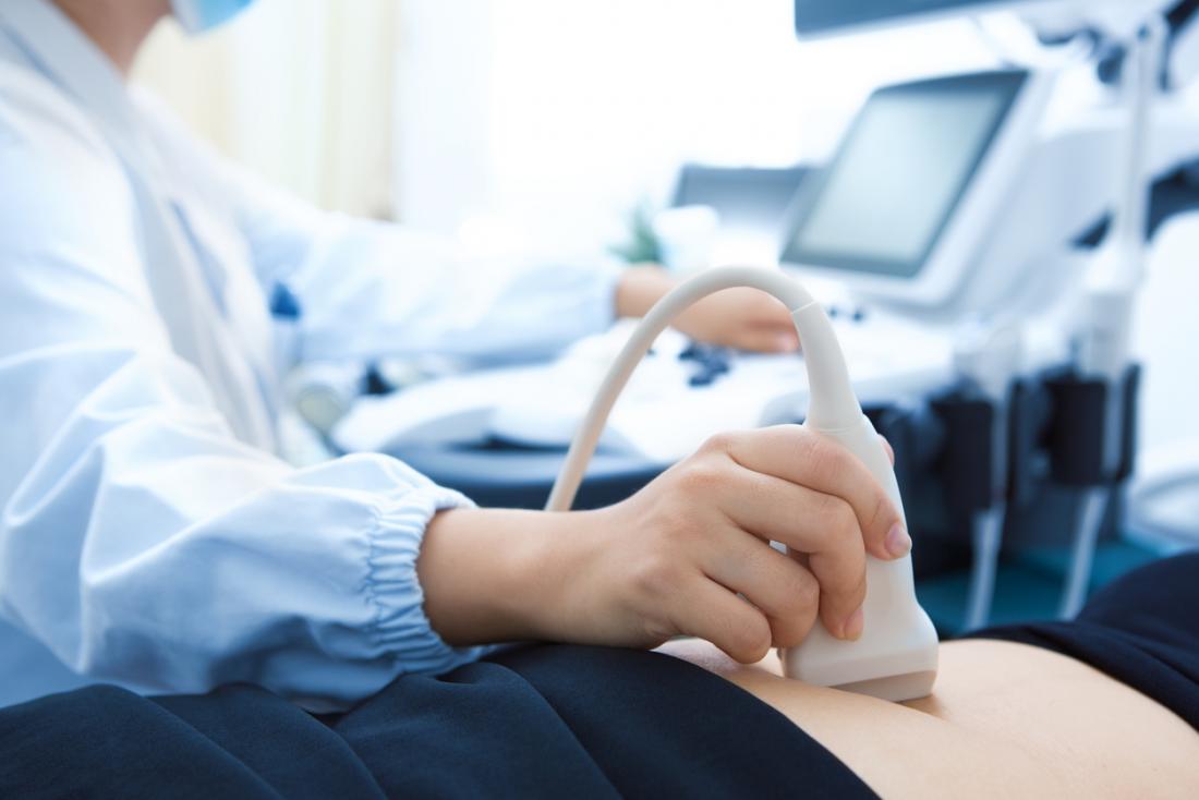
Ultrasound: how does it work?
Ultrasound takes its name from the Greek êchô , which means “sound” and graphy, or graphein in Greek, which means “to write”.
Ultrasound is one of the best-known imaging techniques. It is based on the properties of ultrasound, or sound waves, which are similar to vibrations, not audible to the human ear.
These ultrasounds are emitted in a beam by a probe and they spread out in a straight line. But their trajectory can be deviated when they reach a denser or more rigid medium. The encounter with these “obstacles” creates echoes which reflect the walls of the organs or the tissues. Using a computer, the echoes are converted into a cross-sectional and moving image of the region being examined. The morphology of the zone thus observable can then be analyzed.
This technique can also be associated with the use of a ” radar ” which allows the study of the vessels (doppler), as is the case for cardiac echo-doppler, for example.
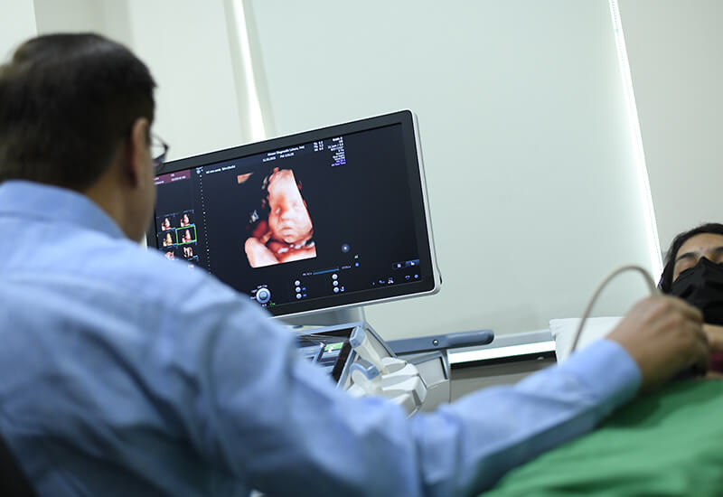
When is an ultrasound performed?
Ultrasound is primarily used to visualize the morphology (size, shape, etc.) of organs, to know their evolution or good development, their location, etc.
This therefore makes it possible to carry out disease diagnoses , to ensure the monitoring of certain pathologies (evolution of cancers , endometriosis , etc.) or to guide the doctor in the context of certain surgical operations . We will not deal with this last aspect, which is more interventional, in this article.
Abdominopelvic ultrasound
The abdomino-pelvic ultrasound (or prostate if it concerns only the prostate) makes it possible to observe the organs of the abdomen and pelvis (uterus, ovaries, prostate and seminal vesicles or liver, gallbladder, kidneys and bladder).
It is useful for diagnosing pathologies that can affect these organs, such as the presence of ovarian cysts or endometriosis within the female reproductive system, or stones in the kidneys, or finally detecting infections of the gallbladder (cholecystitis) or kidneys for example (pyelonephritis).
And it is also known to help monitor the pregnancy of pregnant women and ensure that the fetus is developing well and that there are no complications (miscarriage, ectopic pregnancy, etc.).
Two techniques are used. The organs can be observed through a probe placed on the skin of the belly. Ultrasound is called transcutaneous. In case of difficulties in benefiting from sufficiently clear and analyzable images, ultrasound is performed by endocavitary or endorectal route (for example for prostate ultrasound). The probe is then introduced inside the body (via the vagina or the rectum) to be as close as possible to the organs to be observed.
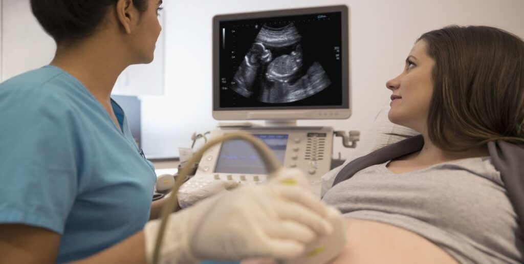
Cardiac echo-doppler
Cardiac echo-doppler is useful if there is a suspicion of cardiovascular disease, it makes it possible to observe several parts in motion. First of all, from the heart (cavities, valves…), but also from the vessels in charge of transporting blood to the heart and to the other organs of the body (this is the case of the aorta and the pulmonary arteries) and finally blood flow, to know how fast and in what direction the blood flows through the vessels. It is therefore used to diagnose diseases, malformations , heart failure ,arterial hypertension … It is also used to ensure follow -up after taking so-called “cardiotoxic” drugs.
It is performed by circulating a probe placed on the thorax and called “transthoracic” (ETT).
Kidney and bladder ultrasound
To explore the kidneys and the bladder, it is the kidney and bladder ultrasound that must be relied upon. It can be prescribed if the patient suffers from abdominal pain or if he is subject to urinary disorders (Presence of blood, pain). It can detect cysts, locate obstruction in the kidneys or stones for example.
Ultrasound in case of tumor
Finally, in the case of a tumor, ultrasound has the function of helping to differentiate between a cyst made up of liquid and a solid tumor, or even to know in which other areas or organs the cancer has developed. For example, thyroid cancer is asymptomatic before leading to potential lumps, difficulty breathing or swallowing. Ultrasound of the thyroid, making it possible to analyze its structure, the size of the gland, and the existence of nodules, is essential in case of doubt.
How is an ultrasound performed?
It can be practiced in many places (hospital, radiology practice, doctor’s office, etc.), as long as the room is sufficiently dark, for good visibility of the images. It does not last long, on average, 15 to 30 minutes and is ambulatory (no need to stay overnight). It can be carried out by a doctor specializing in the organ to be analyzed ( cardiologist , gynecologist, etc.), a radiologist or, in certain cases, by other health professionals.
Before the ultrasound, procedures may be required to make the organ easier to examine. In some cases, a laxative will need to be taken first. It will sometimes be necessary to come on an empty stomach, without drinking or eating, or on the contrary to drink large quantities of water.
An assessment, relating to the medical history, current illnesses and medication taken, is carried out before the ultrasound. For the ultrasound itself, the patient is seated or lying down and may be asked to hold their breath. A probe, previously covered with an ultrasound-conducting gel, is positioned on the area to be analyzed (skin or by insertion into an orifice to be in direct contact with the organ). Connected to the computer, it makes it possible to transcribe moving images of the observed area using ultrasound. Some images are recorded so that the doctor can better analyze the morphology of the organs and in particular take measures. The results are generally communicated and explained after the examination, and formalized in a report, given to the patient and sometimes to the prescribing doctor.
If he deems it necessary, the doctor can recommend additional tests.

Ultrasound, is it painful?
Ultrasounds are not X-rays. The examination does not present any pain, danger or complications for the patient.
However, the pressure of the probe can be unpleasant if the area is weakened or in pain.
What documents to bring for an ultrasound?
If you are required to have an ultrasound, do not hesitate to ask your doctor’s office for the information you will need to provide on D-Day.
In particular, remember to bring your prescription, your carte Vitale, your mutual insurance card, and any other important element of your medical file (Letters from the doctor, results of examinations, reports, old x-rays, ultrasounds, etc.).
What if you get a second opinion?
Have you received ultrasound results, discussed them with your doctor, but still have questions about the diagnosis made and the procedure to follow? A second opinion from a doctor specializing in your illness may be useful to reassure you.


buy synthroid without prescription
Ridiculous quest there. What happened after? Good luck!
Read now. Read now.
https://canadianfast.online/# canadian online drugstore
Read information now. Drug information.
I could not refrain from commenting. Well
written!
order lasix 40 mg
azithromycin 500mg tab cost
Keep on working, great job!
двигатели weka
Read information now. Some are medicines that help people when doctors prescribe.
the canadian drugstore
Long-Term Effects. Commonly Used Drugs Charts.
phenergan tablets
cipro 590 mg
Hey there! Someone in my Facebook group shared this site with us so
I came to give it a look. I’m definitely loving the information. I’m bookmarking and will be tweeting this to
my followers! Wonderful blog and brilliant design and style.
diflucan tablets
Thanks for finally talking about > Everything you need to
know about ultrasound – LabUncle < Loved it!
albenza cost
Hey There. I found your blog the use of msn. This is a really neatly written article.
I will make sure to bookmark it and return to
read extra of your useful information. Thank you for the post.
I will definitely return.
Thanks to my father who stated to me concerning this webpage, this web site is really amazing.
albendazole online uk
steam desktop authenticator
Hey there! Do you know if they make any plugins to protect against hackers?
I’m kinda paranoid about losing everything I’ve worked hard on. Any suggestions?
kamagra 100mg oral jelly ebay
You have brought up a very fantastic details, appreciate it for the post.
Review my blog … Viltrex Powflex Male Enhancement
Hey there are using WordPress for your blog platform?
I’m new to the blog world but I’m trying to get started and
create my own. Do you require any html coding expertise to make your own blog?
Any help would be really appreciated!
where to order prozac a prescription
lasixonline
Pills information sheet. Cautions.
cephalexin buy
Actual information about medication. Get here.
Fantastic post but I was wondering if you
could write a litte more on this subject? I’d be very thankful if you
could elaborate a little bit further. Bless you!
viagra price in india
Very shortly this site will be famous among all blogging and site-building users, due to it’s nice posts
Greetings! This is my first comment here so I just
wanted to give a quick shout out and say I genuinely enjoy reading
your blog posts. Can you suggest any other blogs/websites/forums that cover the same subjects?
Thanks for your time!
Here is my web blog: buy ozempic mexico
fluoxetine 15 mg for sale
buy propecia online no rx
Thanks , I’ve recently been searching for information about this subject for
a long time and yours is the greatest I’ve came upon so far.
However, what about the conclusion? Are you sure concerning the source?
Also visit my blog USA Script Helpers: Affordable Prescription Drugs Canada
It’s very effortless to find out any matter on net as compared
to books, as I found this post at this web site.
Comprehensive side effect and adverse reaction information. п»їMedicament prescribing information.
canada pharmacy online legit
Read information now. drug information and news for professionals and consumers.
Деградация
Психология способностей
Сатира
Оркестровая яма
Художественный образ
Раздражитель
Концепция
Черта характера
lisinopril 240
Ширма
Карнавал
Cautions. Get information now.
https://canadianfast.com/# pain meds without written prescription
Generic Name. safe and effective drugs are available.
where can i get gabapentin
I discovered most gentle stains came out shortly and with out an excessive amount of effort on my half, however I did spend extra time on more durable spots. As a result, it’s straightforward for smaller objects to get misplaced; I usually have to spend time looking for my lipstick or hand sanitizer. This spot cleaner actually isn’t the sexiest buy I’ve made for our new house, however it’s one that may keep it looking contemporary and clean for longer. Investing in a spot cleaner seemed like a no-brainer, and I’m glad I went with Bissell’s SpotClean Pet Professional. It’s really easy to simply seize every thing irrespective of where it’s that I’m going. The SpotClean’s handle has a button that allows you to control the amount of answer it sprays out, and whereas it’s operating, it’s continually sucking up excess water, filling up a separate tank with the dirty bits. I’ve had it for a number of months now, and it’s earned its place as my new everyday bag.
my web site https://www.kogumahome.com/hokkori-blog/2017/06/10/hokkori-diary-0-6/
bactrim over the counter