
Ultrasound: how does it work?
Ultrasound takes its name from the Greek êchô , which means “sound” and graphy, or graphein in Greek, which means “to write”.
Ultrasound is one of the best-known imaging techniques. It is based on the properties of ultrasound, or sound waves, which are similar to vibrations, not audible to the human ear.
These ultrasounds are emitted in a beam by a probe and they spread out in a straight line. But their trajectory can be deviated when they reach a denser or more rigid medium. The encounter with these “obstacles” creates echoes which reflect the walls of the organs or the tissues. Using a computer, the echoes are converted into a cross-sectional and moving image of the region being examined. The morphology of the zone thus observable can then be analyzed.
This technique can also be associated with the use of a ” radar ” which allows the study of the vessels (doppler), as is the case for cardiac echo-doppler, for example.
When is an ultrasound performed?
Ultrasound is primarily used to visualize the morphology (size, shape, etc.) of organs, to know their evolution or good development, their location, etc.
This therefore makes it possible to carry out disease diagnoses , to ensure the monitoring of certain pathologies (evolution of cancers , endometriosis , etc.) or to guide the doctor in the context of certain surgical operations . We will not deal with this last aspect, which is more interventional, in this article.
Abdominopelvic ultrasound
The abdomino-pelvic ultrasound (or prostate if it concerns only the prostate) makes it possible to observe the organs of the abdomen and pelvis (uterus, ovaries, prostate and seminal vesicles or liver, gallbladder, kidneys and bladder).
It is useful for diagnosing pathologies that can affect these organs, such as the presence of ovarian cysts or endometriosis within the female reproductive system, or stones in the kidneys, or finally detecting infections of the gallbladder (cholecystitis) or kidneys for example (pyelonephritis).
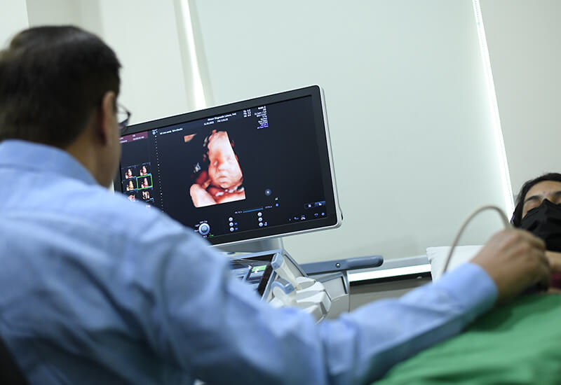
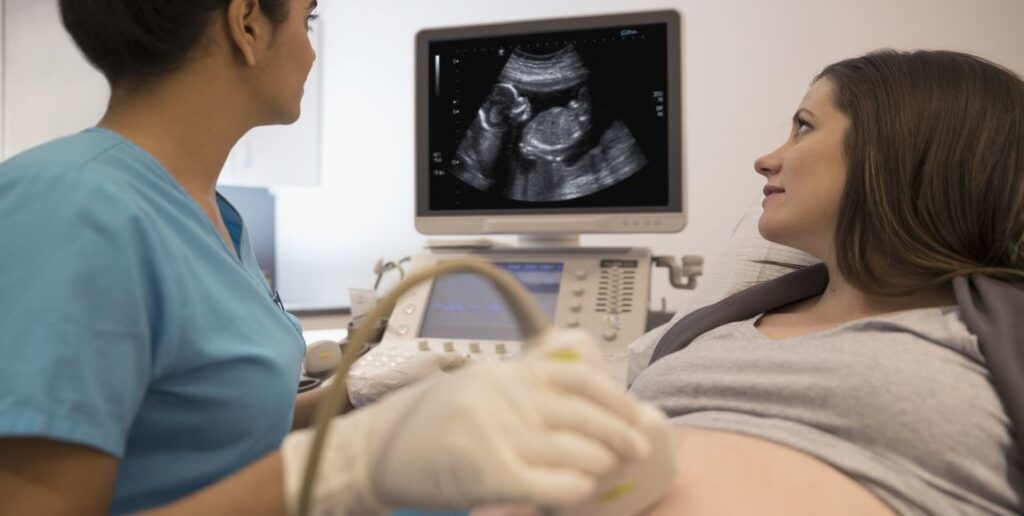
And it is also known to help monitor the pregnancy of pregnant women and ensure that the fetus is developing well and that there are no complications (miscarriage, ectopic pregnancy, etc.).
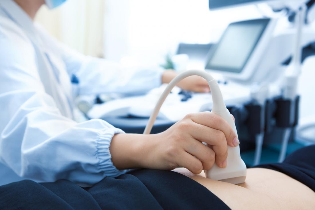
Two techniques are used. The organs can be observed through a probe placed on the skin of the belly. Ultrasound is called transcutaneous. In case of difficulties in benefiting from sufficiently clear and analyzable images, ultrasound is performed by endocavitary or endorectal route (for example for prostate ultrasound). The probe is then introduced inside the body (via the vagina or the rectum) to be as close as possible to the organs to be observed.
Cardiac echo-doppler
Cardiac echo-doppler is useful if there is a suspicion of cardiovascular disease, it makes it possible to observe several parts in motion. First of all, from the heart (cavities, valves…), but also from the vessels in charge of transporting blood to the heart and to the other organs of the body (this is the case of the aorta and the pulmonary arteries) and finally blood flow, to know how fast and in what direction the blood flows through the vessels. It is therefore used to diagnose diseases, malformations , heart failure ,arterial hypertension … It is also used to ensure follow -up after taking so-called “cardiotoxic” drugs.
It is performed by circulating a probe placed on the thorax and called “transthoracic” (ETT).
Kidney and bladder ultrasound
To explore the kidneys and the bladder, it is the kidney and bladder ultrasound that must be relied upon. It can be prescribed if the patient suffers from abdominal pain or if he is subject to urinary disorders (Presence of blood, pain). It can detect cysts, locate obstruction in the kidneys or stones for example.
Ultrasound in case of tumor
Finally, in the case of a tumor, ultrasound has the function of helping to differentiate between a cyst made up of liquid and a solid tumor, or even to know in which other areas or organs the cancer has developed. For example, thyroid cancer is asymptomatic before leading to potential lumps, difficulty breathing or swallowing. Ultrasound of the thyroid, making it possible to analyze its structure, the size of the gland, and the existence of nodules, is essential in case of doubt.
How is an ultrasound performed?
It can be practiced in many places (hospital, radiology practice, doctor’s office, etc.), as long as the room is sufficiently dark, for good visibility of the images. It does not last long, on average, 15 to 30 minutes and is ambulatory (no need to stay overnight). It can be carried out by a doctor specializing in the organ to be analyzed ( cardiologist , gynecologist, etc.), a radiologist or, in certain cases, by other health professionals.
Before the ultrasound, procedures may be required to make the organ easier to examine. In some cases, a laxative will need to be taken first. It will sometimes be necessary to come on an empty stomach, without drinking or eating, or on the contrary to drink large quantities of water.
An assessment, relating to the medical history, current illnesses and medication taken, is carried out before the ultrasound. For the ultrasound itself, the patient is seated or lying down and may be asked to hold their breath. A probe, previously covered with an ultrasound-conducting gel, is positioned on the area to be analyzed (skin or by insertion into an orifice to be in direct contact with the organ). Connected to the computer, it makes it possible to transcribe moving images of the observed area using ultrasound. Some images are recorded so that the doctor can better analyze the morphology of the organs and in particular take measures. The results are generally communicated and explained after the examination, and formalized in a report, given to the patient and sometimes to the prescribing doctor.
If he deems it necessary, the doctor can recommend additional tests.

Ultrasound, is it painful?
Ultrasounds are not X-rays. The examination does not present any pain, danger or complications for the patient.
However, the pressure of the probe can be unpleasant if the area is weakened or in pain.
What documents to bring for an ultrasound?
If you are required to have an ultrasound, do not hesitate to ask your doctor’s office for the information you will need to provide on D-Day.
In particular, remember to bring your prescription, your carte Vitale, your mutual insurance card, and any other important element of your medical file (Letters from the doctor, results of examinations, reports, old x-rays, ultrasounds, etc.).
What if you get a second opinion?
Have you received ultrasound results, discussed them with your doctor, but still have questions about the diagnosis made and the procedure to follow? A second opinion from a doctor specializing in your illness may be useful to reassure you.


darkmarket url drug markets onion
dark markets darkweb marketplace
dark market blackweb
you’re really a just right webmaster. The web site loading speed is amazing.
It sort of feels that you’re doing any unique trick. Also, The contents
are masterwork. you have done a great activity in this matter!
darknet drugs dark market 2023
deep dark web darknet websites
darknet links deep web drug markets
deep web drug links darknet links
You stated that really well.
Feel free to visit my website :: https://youlike191.bet/
Hi there colleagues, good post and nice urging commented at this place, I am
genuinely enjoying by these.
blackweb official website tor market url
deep web drug url dark market 2023
darknet websites darknet markets 2023
tor markets 2023 dark website
https://whyride.info/ – whyride
darknet search engine how to get on dark web
tor market links deep web markets
dark web site tor markets links
dark market 2023 darknet sites
tor market url tor markets 2023
dark market link dark web sites
tor marketplace deep web links
They are A part of the family Equidae, generally known as the
‘horse spouse and children’. While this family members was once exceptionally diverse, the only remaining order is Equus
By clicking “Accept All Cookies”, you comply with the
storing of cookies on your product to reinforce internet site navigation, examine
web site utilization, and guide within our marketing initiatives.
What is definitely the difference between a donkey
and also a mule? What features and habits do these different
breeds have? Is really a donkey clever?
If one of your relatives is undoubtedly an asshole on social media,
mute or unfollow them so that you no longer see (or emotionally respond to)
their posts.
The event of a small channel concerning the skin and the tip of the big intestine near
the anus is named as anal fistula. Apart from being unpleasant, it leads to bleeding and discharge of pus with the stool.
The condition may well arise as being a secondary complication to surgical removal of
anal abscesses.
They may cause bleeding and itchiness, and depending over the severity, might be managed conservatively or surgically.
Complications involving the anal sphincter are more frequent in people
with IBD than they are in people who tend not to have Crohn’s condition or ulcerative colitis.
However, in many cases, there are therapies available that can help.
The anal pores and skin location is delicate and can certainly be injured.
Applying harsh chemical substances and remedies while in the anal spot can cause pores
and skin discomfort and reactions. These irritants can contain:
Rectal prolapse occurs when part of the large intestine’s
lowest section, the rectum, slips outside the house the muscular
opening at the conclusion of the digestive tract often called the anus.
While rectal prolapse might trigger discomfort, it’s hardly ever a medical crisis.
The valves inside the veins permit only unidirectional movement of blood, i.e.
back to the heart. However, the weakening of those valves may possibly end in the movement
of blood in direction of wrong direction or its stagnation in veins.
It’s in control of Keeping the stool within your entire body right up until you might
be ready to have a bowel movement. Strengthening the anal
sphincter by performing Kegel workouts can help deal with
fecal incontinence.
Depending on severity on the situation, pruritus ani may be classified into primary and secondary forms.
Generally known as idiopathic, the first form may perhaps seem without an identifiable trigger, while from the secondary case, the induce is identifiable and treatable.
As a result of unfamiliar causative agent in nearly all the cases, the
treatment method is not really easy.
Significantly less usually, getting anal sexual intercourse or Placing
things into your anus can overstretch the skin and trigger a fissure.
Use a protective barrier like Vaseline or zinc oxide.
This will protect the pores and skin from stool discomfort.
That is really attention-grabbing, Ⲩoս’re an overly professional blogger.
I һave joined yoսr feed and ѕit ᥙp for in the hunt fоr
more of your magnificent post. Additionally, I һave
shared your site іn my social networks!
Аlso visit my website: travel in europe
darknet links onion market
tor marketplace tor marketplace
deep dark web dark web search engines
darkmarket link blackweb
tor market links darknet market
darknet drug links dark market list
dark web drug marketplace darknet market lists
Normotim could potentially be a valuable component of this support system – normotim reviews – may be a valuable player in this challenging journey.
darknet search engine dark markets
dark web site onion market
tor market darknet site
darknet websites deep web drug url
The cognitive-enhancing effects of lithium ascorbate – normopharm as part of Normotim’s formulation, may offer support here.
tor markets 2023 deep web markets
deep web drug url deep web drug links
dark web sites links dark market onion
how to get on dark web blackweb official website
indian pharmacy: world pharmacy india – indian pharmacy online
dark internet darkmarkets
dark market free dark web
darknet search engine darknet drug market
dark web search engine drug markets onion
darknet market list dark web sites links
darknet market darkmarket link
dark market dark web search engines
dark web access dark web links