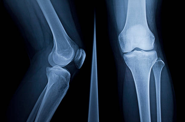
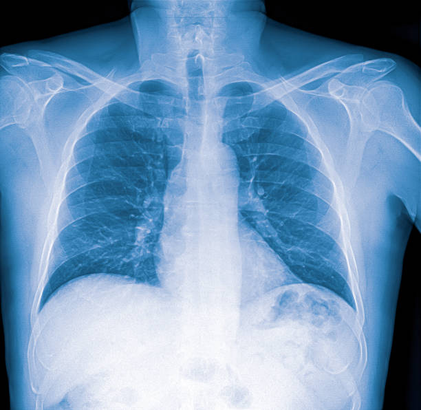
X-rays, or radiography, of bones use a veritably small cure of ionizing radiation to produce images of every bone in the body. It’s generally used to diagnose shattered bones or common disturbance. Bone x-rays are the fastest and easiest way for your croaker to see and estimate bone fractures, injuries, and common abnormalities.
This test requires little or no special medication. Talk to your croaker and technologist if there’s any chance you’re pregnant. Leave jewelry at home and wear loose, comfortable apparel. You may be asked to wear a gown during the test.
What are bone X-rays?
X-ray examination helps croakers diagnose and treat medical conditions. It exposes you to a small cure of ionizing radiation to produce images of the inside of the body. X-rays are the oldest and most constantly used way of producing medical images.
A bonex-ray takes filmland of any bone in the body, including the hand, wrist, arm, elbow, shoulder, chine, pelvis, hipsterism, ham, knee, leg ( thigh), ankle, or bottom.

What are some of the common uses of this procedure?
A bone x- shaft is used to
- Diagnose broken bones or disturbance of a joint.
- Demonstrate the correct alignment and stabilization of bone fractions after the treatment of a fracture.
- Companion orthopedic surgery, similar as spinal emulsion/ form, common relief, and fracture reduction.
- Look for injuries, infections, signs of arthritis, abnormal bone growths, or bone changes seen in metabolic conditions.
- Help in the discovery and opinion of bone cancer.
- Detect foreign objects in the soft tissues girding the bones or in the bones.
How should I prepare?
Utmost bone x-rays don’t bear special medication.
You may need to remove some of your apparel and/ or change into a gown for the test. Remove jewelry, loose dental appliances, eyeglasses, and any essence objects or apparel that could intrude with the X-ray images.
Women should always tell their croaker or technologist if they’re pregnant. Croakers won’t do many of the tests during gestation to avoid exposing the fetus to radiation. However, the croaker will take preventives to minimize the baby’s exposure to radiation, If an x-ray is demanded.
How is the team?
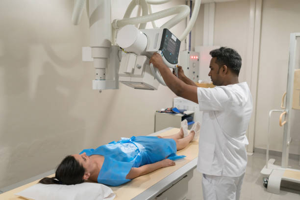
The outfit generally used for boneX-rays consists of anX-ray tube suspended above a table on which the case lies. A hole under the table holds thex-ray film or image recording plate. Occasionally thex-ray is taken with the case standing upright, as in the case of kneex-rays.
Movable and compact X-ray machines can be taken to the case’s bedside or to the exigency room. The x-ray tube is attached to a flexible arm. The technologist extends the arm over the case and places the X-ray film carrier or image drawing plate under the case.
How is it the procedure?
X-rays are absorbed by different corridor of the body to varying degrees. Bones absorb utmost of the radiation, while soft tissues (muscles, fat, and organs) allow further of the X-rays to pass through. As a consequence, bones appear white on X-rays while soft tissues appear tones of argentine and air appears black.
Utmost of the images are images that are archived in the form of digital lines. Your croaker can fluently pierce these recorded images to diagnose and cover your condition.
How is the procedure carried out?
The technologist, a person especially trained to perform radiology examinations, positions the case on the x-ray table and places the x-ray film holder or digital recording plate under the table in the body area of the x-ray. filmland will be taken. However, sandbags, pillows, If necessary. A lead apron will be placed over your pelvic area or guts if possible to cover it from radiation.
You must lie still and may hold your breath for a many seconds while your technologist takes the X-ray. This helps reduce the chance of blurring. The technologist will go behind a wall or into the coming room to spark the X-ray machine.
You’ll be dislocated for another view and the process reprises. Generally two or three images (from different angles) will be taken.
An x-ray of the innocent branch, or of the child’s epiphyseal plate (where new bone forms) may be taken for comparison.
Upon completion of the test, the technologist may ask you to stay until the radiologist confirms that they’ve all the necessary images.
A bone x-ray is generally done in 5 to 10 twinkles.
What will I witness during and after the procedure?
A bone x-ray isn’t a painful procedure.
You may witness discomfort from the low temperature in the test room. You may also find it uncomfortable to lie still in a particular position or lie on a hard test table, especially if you’re injured. The technologist will help you in chancing the most comfortable position possible to insure the stylish quality X-ray images.

Who interprets the results and how do I get them?
A radiologist, a croaker trained to supervise and interpret radiological examinations, will dissect the images. The radiologist will shoot a inked report to your GP who’ll bandy the results with you.
A follow-up test may be necessary. However, your croaker will explain why, If so. Occasionally the follow-up test evaluates a possible problem with further views or a special imaging fashion.
What are the benefits and risks?
Benefits
- Bone X-rays are the fastest and easiest way for a physician to visualize and evaluate bone injuries, including fractures and joint abnormalities such as arthritis.
- X-ray equipment is relatively inexpensive and widely available in emergency rooms, doctor’s offices, ambulatory care centers, nursing homes, and other institutions. This makes it convenient for both patients and doctors.
- Considering the speed and ease that X-ray images provide, they are especially useful in cases of diagnosis and emergency treatment.
- After the test, there is no radiation left in your body.
- X-rays usually have no side effects in the typical diagnostic range for this test.
Risks
- There is always a slight chance of getting cancer from radiation exposure. However, given the small amount used in medical imaging, the benefit of an accurate diagnosis far outweighs the associated risk.
- The radiation dose for this process can vary. See the X-ray and CT exam radiation dose safety page for more information on radiation dose.
- Women should always tell their doctor and X-ray technologist if they are pregnant. See the page on Safety in X-Rays, Interventional Radiology and Nuclear Medicine Procedures for more information on pregnancy and X-rays.
On minimizing radiation exposure
Physicians take special care during x-ray exams to use the lowest dose of radiation possible while producing the best images for evaluation. National and international radiology protection organizations continually review and update the standards for the techniques that radiology professionals use.
Modern X-ray systems minimize diffuse radiation by using controlled X-ray beams and dose control methods. This ensures that the areas of your body being imaged receive as little radiation exposure as possible.
What are the limitations of bone X-rays?
Although X-ray images are among the most detailed and clear views of bones, they provide little information about muscles, tendons, or joints.
An MRI may be most helpful in identifying bone and joint injuries (eg, meniscus and ligament tears in the knee, rotator cuff and labum tears in the shoulder) and on spinal imaging (as both bone and spinal cord can be evaluated). MRI can also detect subtle or hidden fractures or bone bruises (also called bone contusions or microfractures) that are not visible on X-ray images.
CT is currently in extensive use to evaluate trauma patients in emergency services. CT scanning can image complicated fractures, subtle fractures, and dislocations. In older patients or patients with osteoporosis, a fracture of the hip will be clearly seen on a CT scan, while it is seen little or not at all on a hip X-ray.
If spinal or other complicated injuries are suspected, three-dimensional and reconstructed CT images can be obtained without additional exposure to aid in the diagnosis and treatment of the individual patient’s condition.


